The nervous system is the most complex and, at the same time, the most delicate biological structure in the human body. It consists of billions of neurons and trillions of synapses that shape our behavior, memory, emotions, and cognition. In neuroscience, many concepts are abstract, such as electrochemical transport, action potentials, and communication networks. Without visualization, these concepts are often unimaginable and difficult to grasp for most audiences.
The nervous system illustration bridges the gap between abstract subjects and tangible understanding. It is crucial for simplifying and translating the complexity of nervous system concepts. From early hand-drawn sketches to modern 3D renderings, this article explores the creation and methods of neuron and nervous system illustrations. By combining scientific accuracy with artistic techniques, such illustrations enhance learning, medical training, and public awareness.
A Brief History of Nervous System Illustration
The field of neural illustration has deep roots. Nobel Prize winners such as Santiago Ramón y Cajal revolutionized neuroscience through meticulous drawings of neurons using the Golgi staining method. His “neuron doctrine” illustrations portrayed neurons as distinct, tree-like cells, laying the foundation for modern neuroscience art. Since pencil drawings in textbooks were replaced by digital tools in the 20th century, medical models now incorporate AI to provide unprecedented detail and interactivity.
Levels of Neural Illustration
There are several levels in neural illustration, each containing its own specific details and style:
- Molecular level: including ion channels, neurotransmitters, and receptors.
- Cellular level: focusing on neurons and other related neural cells.
- Tissue and organ level: illustrating brain regions, spinal nerves, and peripheral networks.
- Systemic level: demonstrating interactions between the brain, spinal nerves, and peripheral nerves in a comprehensive map.
These levels build upon each other, allowing illustrators to zoom in or out for context.
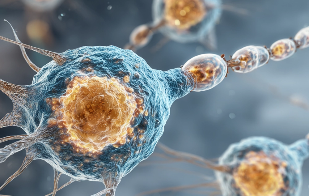
Neuron Structures
Every neuron consists of four main parts:
- The soma
- Dendrites
- Axon
- Synapse


Forming an overall tree-like structure with a trunk and multiple branches. These designs are not only visually attractive but also convey the concept of extended connections.
Based on their functions and locations, neurons exhibit various shapes, such as:
- Multipolar (many dendrites)
- Bipolar (one dendrite and one axon)
- Unipolar (a single process)
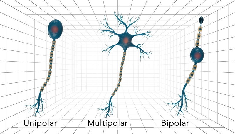
The different morphologies of these types should be considered in a precise scientific illustration. The myelin sheath covers axons like the insulation of electrical wires and increases signal velocity. It is usually depicted as white or semi-transparent rings surrounding axons at regular intervals.
Additionally, neurons are classified into several types based on the morphology of their soma and the pattern of their appendages, such as:
- Pyramidal
- Stellate
- Spindle-shaped
- Spherical
- Pear-shaped neurons
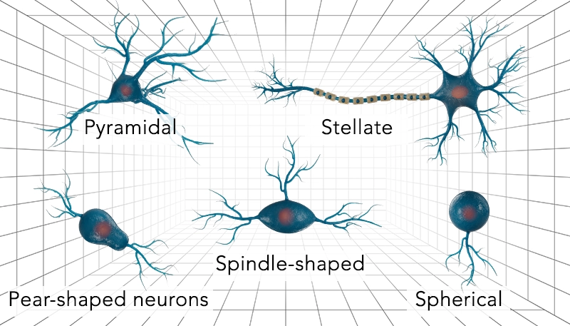
These variations in structure result in distinct functions and properties across different parts of the nervous system.
Other Cells in the Nervous System
Aside from messenger neurons, several glial cells play key roles in the performance and maintenance of the nervous system:
• Astrocytes:
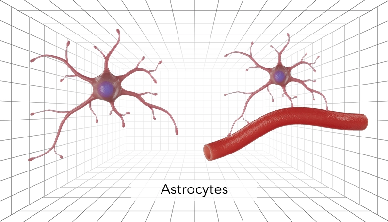
Provide neurons with nutrition and have a star-shaped appearance (hence the name). These cells consist of a central body with several slender extensions and are designed to appear semi-transparent with soft colors, sometimes glowing to emphasize their supporting role. Their extensions can also wrap around synapses and capillaries.
• Oligodendrocytes and Schwann cells
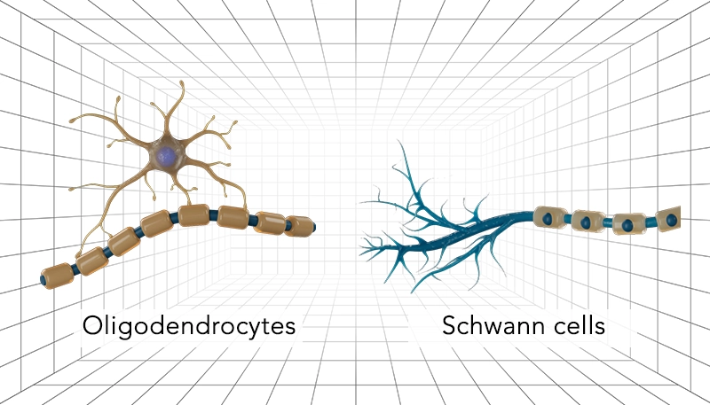
Create the myelin sheath. These two types of cells share the same function but are found in different regions. Oligodendrocytes are present in the central nervous system, while Schwann cells are located in the peripheral nervous system. They are usually visualized as small, round cells in light colors, with the myelin sheath surrounding the axons.
• Microglia
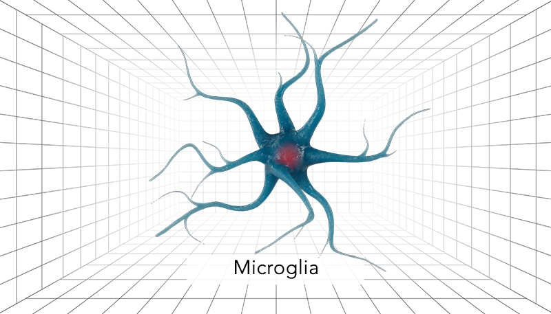
Act as immune cells in the nervous system and are the smallest glial cells. They are illustrated with a dark, small body and irregular, short appendages, sometimes resembling macrophages.
In illustrations, combine these with neurons using varied colors, contrasts, and transparencies for realism and informativeness.
Synapse and Signaling
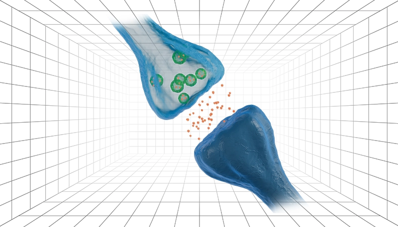
A synapse can be illustrated as two terminal ends positioned close to each other with a shallow cleft between them. In the pre-synaptic terminal, tiny sacs release neurotransmitters into the synaptic space. In digital visualizations, this moment is often depicted using glowing molecules and light effects to make the process more tangible. Neurons transmit signals through two pathways—electrical and chemical—which can be represented by luminous waves, dynamic signals, or shining points.
Neural Networks and Nervous System
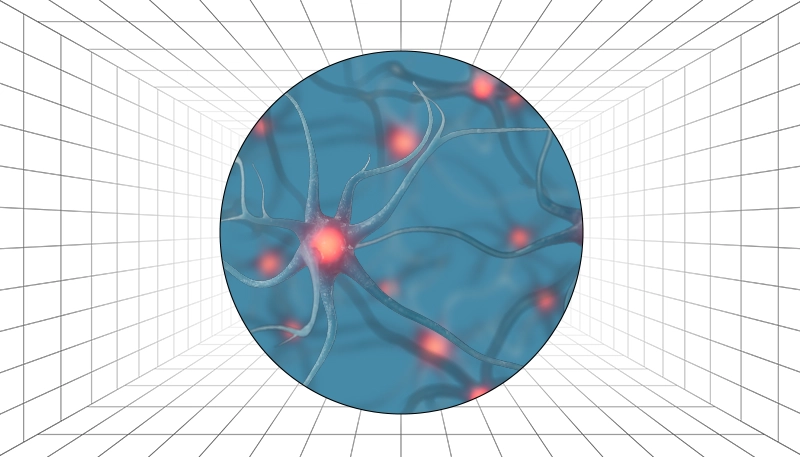
In neural illustrations, individual neurons are often designed as dynamic elements within a vast, interconnected network. Each neuron is positioned to reflect real biological organization—densely packed in certain areas and more dispersed in others—creating a sense of depth and communication across the structure. Subtle variations in color, size, and brightness are used to represent the flow of information and the complexity of neural connectivity.
At the network level, the aim is to illustrate interconnected circuits ranging from simple neuronal pairs to highly complex brain regions. Graphical patterns such as clustered, lattice-based, or organic free-form arrangements can be employed to demonstrate the diverse connectivity of the human brain and its similarity to artificial neural networks. In these visualizations, line thickness, glow intensity, or nodal density may be applied to indicate the strength or direction of information flow.
The nervous system is divided into two major parts: the central nervous system (CNS)—which includes the brain and spinal cord—and the peripheral nervous system (PNS), consisting of all nerves extending beyond the CNS. To demonstrate the brain, simplified 3D models are commonly used, with color coding applied to distinguish different regions such as the cerebrum, cerebellum, and brainstem. Transparent sections, half-cut views, or cross-sectional layers are often incorporated to reveal internal brain structures and pathways.
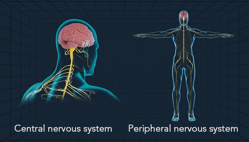
In contrast, illustrations of the peripheral nervous system emphasize the branching pattern of nerves throughout the body. These networks are typically shown as fine, thread-like extensions spreading from the spinal cord to muscles and sensory organs. Using soft gradients or semi-transparent overlays can effectively convey the continuity between central and peripheral components, helping viewers visualize how information travels from the brain to the body and back.
Color Symbolism, Artistic Metaphor
In scientific illustration, color serves a purpose far beyond aesthetics — it carries meaning. Each color can convey specific information about neural activity and function. Cool tones such as blue and cyan often represent calmness, stability, or the flow of information. Warm colors like yellow and gold symbolize energy, transmission, and excitation, while red highlights areas of activity or stimulation. Purple or violet shades may indicate inhibition or suppression within neural circuits. The deliberate contrast between these colors across different regions of the nervous system helps audiences grasp signal direction, intensity, and dynamic interactions.

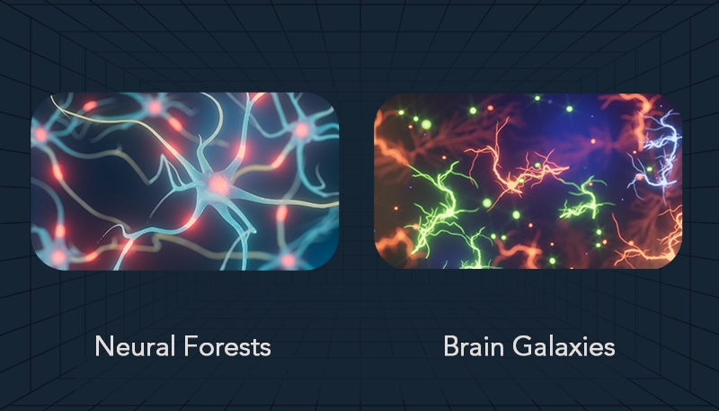
Illustrators frequently use metaphors to make complex scientific concepts more accessible and emotionally engaging. Neurons may be visualized as “neural forests,” networks as “brain galaxies,” or signaling as “lightning.” These artistic parallels transform highly technical information into imagery that feels vivid and relatable.
To create depth and movement in neural illustrations, artists employ techniques such as color gradients, glowing synapses to highlight activity, motion blur to simulate the transmission of impulses, and layers of transparency to represent the complexity of overlapping networks. These elements work together to build dynamic, visually rich scenes that communicate both structure and function.
In 3D design, two main visualization approaches are commonly used. The first involves solid 3D models, in which neural structures have physical volume and tangible form. These models are often used in medical education or anatomical studies, where accuracy, proportion, and spatial relationships are essential. They allow rotation, zooming, and interaction — helping learners perceive how different parts of the nervous system connect in real space.
The second approach relies on light-based 3D representations, where structures are rendered as glowing filaments, particles, or light trails rather than solid forms. This style emphasizes the dynamic and abstract nature of neural activity — ideal for visualizing connectivity, signal transmission, or brain-computer interface concepts. These models evoke a sense of flow and energy rather than anatomy.
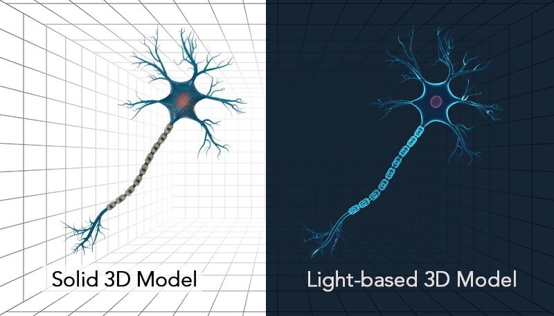
In practice, both methods are valuable: solid models are favored in scientific and medical contexts that require precision, while light-based models dominate in educational media, documentaries, and artistic visualizations. However, light-based visualizations are more frequently used when illustrating neural networks, as they better capture the immaterial, energetic essence of the nervous system and its communication processes.
Who Benefits from Nervous System Illustration?
Nervous system illustrations can help teachers and students simplify complex concepts, assist researchers in visualizing their data, support medical professionals in planning therapies, and aid public health initiatives in raising awareness about neural disorders.
In general, these images are widely used in the following categories:
- Education: In textbooks, online courses, and classroom diagrams to simplify complex anatomy.
- Scientific Research: For visualizing data in journal articles, grant proposals, and connectome studies.
- Medical Training: In simulations for neural disorders, surgical planning, and patient education.
- Public Awareness: Through infographics on brain health and campaigns for mental illness awareness.
- Animations and Videos: For dynamic depictions of signal transmission.
- Art and Media: In neurographic artworks used for aesthetic or therapeutic purposes.
Nervous System Illustration in Covers
This cover art from Nature Neuroscience beautifully blends science and symbolism to illustrate the concept of neural fragility in epilepsy. The image shows a chalice from which flames and smoke rise, transforming into a brain. The orange flames represent fragile epileptogenic neurons—the source of seizures—while the blue smoke symbolizes hyperactive EEG signals. Visually, the transition from fire to smoke to brain represents the process by which unstable neurons lead to widespread neural activity. As a result of the dark background and glowing light effects, epileptic activity is emphasized in a striking way as a metaphor for the delicate balance of the human brain.
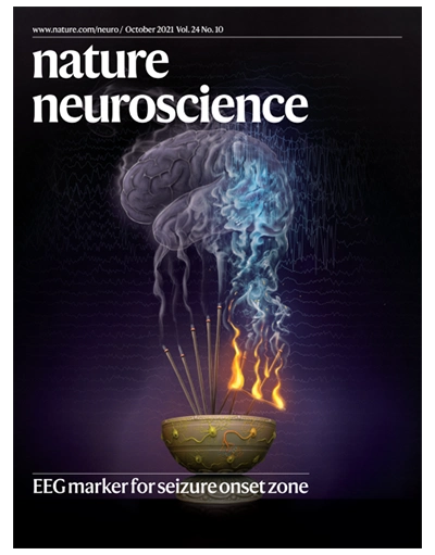

Nature Neuroscience, Volume 24, Issue 10, October 2021 (Cover Created by Shawna Snyder)
This cover art from Advanced Science vividly represents the intersection of molecular design and neural function in neuromorphic technology. The image depicts a stylized synapse — the communication bridge between neurons — with glowing signals symbolizing the transmission of electrical impulses. The surrounding molecular chains illustrate the alkyl side chains of polymers, whose length adjustment is key to enhancing non-volatile properties in synaptic devices. The dynamic colors of blue and purple highlight the flow of ions across the synaptic junction, capturing both the energy and precision of electronic signaling. In general, the artwork demonstrates how fine molecular tuning can mimic the adaptive intelligence of the human brain.
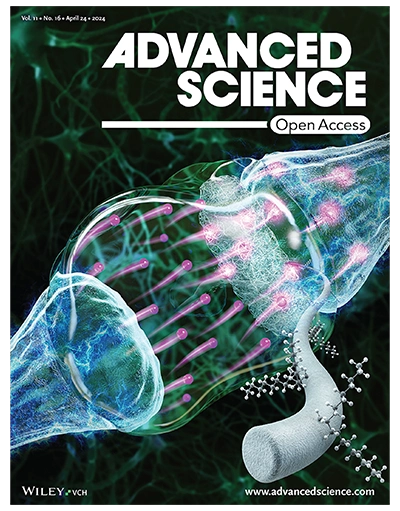
Advanced Science, Volume 11, Issue 16, April 2024 (Cover Created by unknown team)
This cover art from Science Advances combines scientific precision with a visually poetic representation of neural communication and memory formation. A dynamic synapse is depicted in blue and orange tones – where electric energy and molecular interaction meet. The glowing cyan spheres symbolize insulin-like growth factors (IGF1/IGF2), released from the postsynaptic side, illuminating the delicate exchange that drives synaptic plasticity and learning. The interplay between cool blue light and fiery orange proteins captures the balance between growth and energy within the brain’s neural circuits. Artistically, the image conveys motion and life: luminous particles appear to pulse and flow like constellations in a microscopic universe.
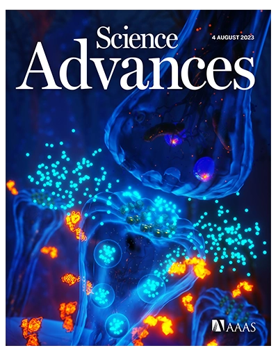
Science Advances, Volume 9, Issue 31, August 2023 (Cover Created by Ella Maru Studio)
This cover artwork for Small magazine beautifully blends science and art to illustrate the intersection of technology and biology. The image shows pink neuron-like structures spreading across a sleek, futuristic surface, connected to shiny metallic nanoprobes. These elements symbolize the advanced sensing platform described in the article — a 3D nanostructured system that records neuronal electrical signals and detects pH changes. A sense of energy and communication is created by the glowing light effects and electric currents, which echo how neurons communicate information. It emphasizes the fusion of organic life and high-tech innovation by using a dark background and bright pink and blue highlights to make the scientific concept visually engaging and dynamic.
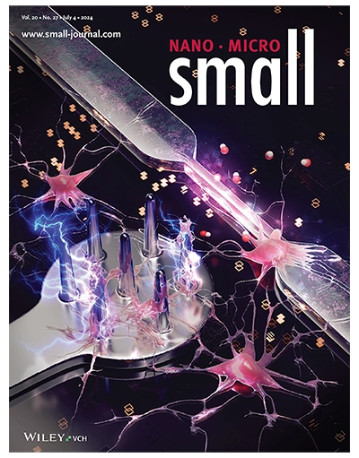
Small, Volume 20, Issue 27, July 2024 (Cover Created by Sciencebrush Studio)
The artwork on this cover beautifully incorporates science and art to represent a new method for studying the brain at the molecular level. In the image, a frozen piece of brain tissue is being sampled, revealing neuronal fibers and synapses in glowing blue tones. With cool colors and smooth textures, the images convey precision, clarity, and the cold environment of cryoelectronic tomography. Symbolizing the process of identifying molecular details in vitrified brain layers, the composition leads the viewer’s eye from the intricate inner structures of the neuron to the cube of frozen tissue.
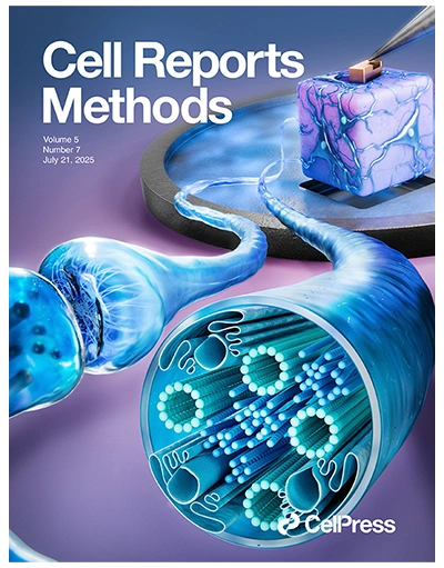
Cell Reports Methods, Volume 5, Issue 7, July 2025 (Cover Created by Inmywork Studio)
This vibrant cover artwork illustrates the fusion of biology and technology through a “neuronal membrane on a chip.” It represents the connection between living systems and bioelectronic devices by depicting a detailed neuron cell membrane with ion channels interacting with an electronic circuit below. The glowing pathways and soft lighting suggest dynamic electrical communication, while the bright colors of lipids and proteins highlight the complexity and vitality of cellular processes. By merging organic neuroscience with advanced electronic platforms, the design explores ion channel behavior in remarkable detail.
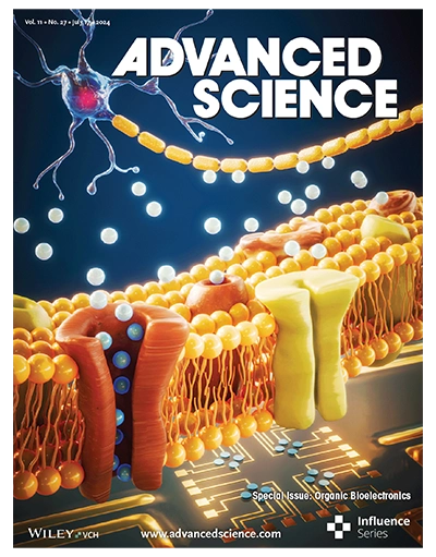
Advanced Science, Volume 11, Issue 27, July 2024 (Cover Created by Inmywork Studio)
Conclusion
Nervous system illustrations fuse science and art, making abstract neurology visible and inspiring. Through structure, color, light, and motion, they reveal brain dynamics for education, research, and communication. As tools evolve, including AI, visualizations grow more immersive, unlocking new insights into our intricate neural world.
At Inmywork Studio, our team of scientific illustrators, 3D designers, and storytellers creates accurate, stunning artworks. We transform complexity into clarity for education, medical visuals, research, or outreach. Visit our website for custom projects.
Recent Journal Cover Designs