Graphene is a 2-dimentional (2D) carbon nanomaterial that is composed of a single layer of carbon atoms, arranged in a hexagonal lattice. Graphene exhibits remarkable properties such as high electrical and thermal conductivity, mechanical strength, and optical transparency. These features make graphene one of the most promising materials in modern applications such as nanoelectronics, biosensing, energy storage, and drug delivery.
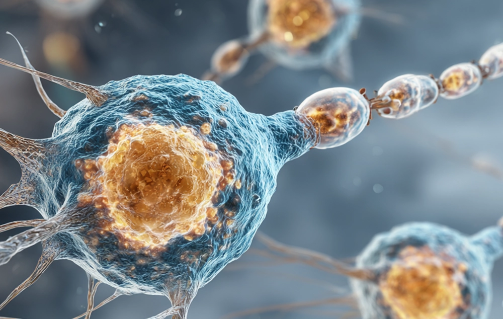
Graphene is atomically thin and it shows complex surface interactions. Therefore, accurate visualization is essential for scientists and engineers to fully understand its structure, properties, and functional modifications. Graphene illustration plays a key role in representing morphology, surface, and its interactions with other nanomaterials or molecules.
Graphene illustration can generally be divided into two categories: experimental methods and digital visualization.
Digital Modeling Methods for Graphene illustration
Digital visualization offers scalable and flexible methods for graphene illustration in scientific publications such as graphical abstracts and cover arts, educational materials, and 3D models. These methods can be used to illustrate both atomic-level structure and macroscopic forms of the graphene-based material.
-
Atomic Lattice Models
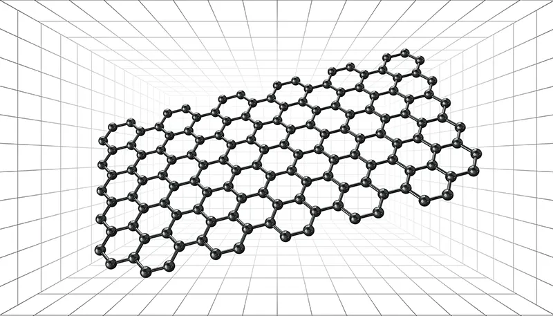
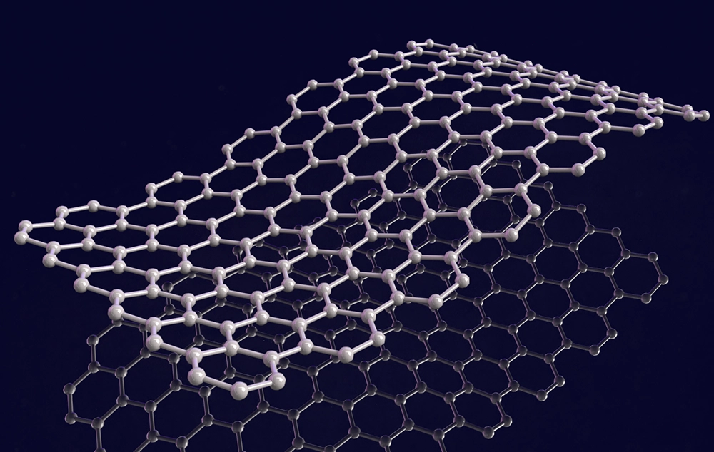
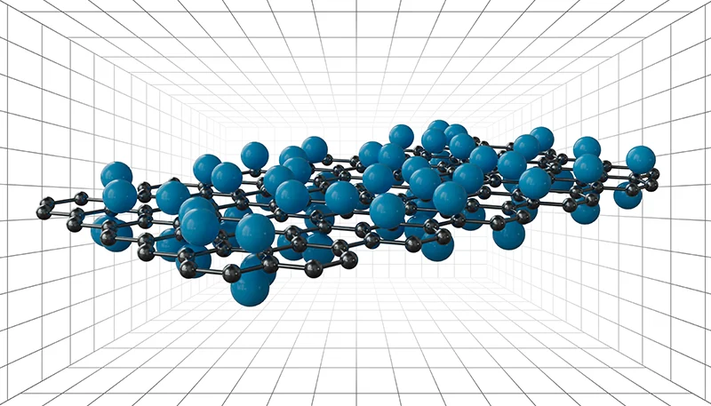
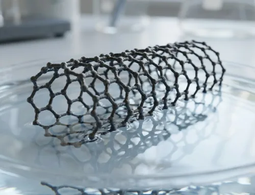
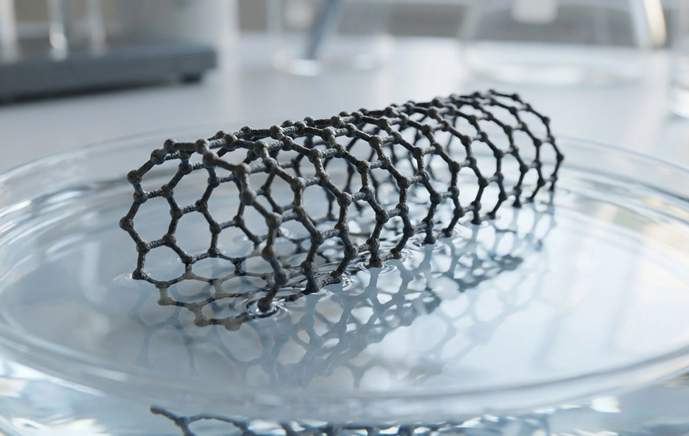
Graphene sheets are mainly shown as a hexagonal honeycomb lattice, which represents carbon atoms connected to each other by sp2 bonds. These models exhibit the perfect symmetry of the graphene structure.
-
Layered Sheet Models
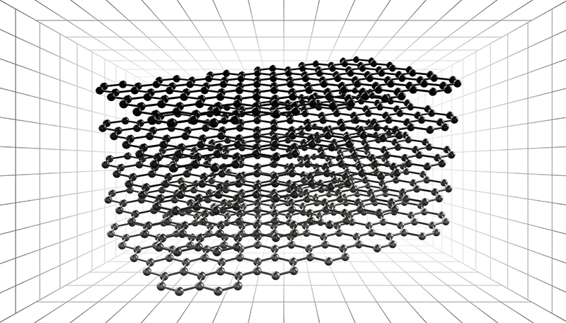
If the intention is to illustrate a few-layer graphene or graphene oxide, stacked transparent sheets can be used. In these models, color gradients or opacity changes indicate the number of layers and the degree of oxidation or reduction of the graphene sheets.
-
Curved Surfaces
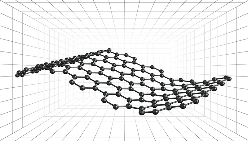
Graphene has a flexible structure and a high surface area. To illustrate these features and visualize the strain and surface visualization, digital models sometimes show folded, wrinkled, or curved graphene surfaces.
-
Composites or Functionalized Structures
Graphene can be combined with a variety of polymers, nanoparticles, or molecules. This type of graphene is often exhibited as a hybrid or embedded network to show interactions between graphene sheets and other components. Functionalized graphene or graphene-based composites are useful in drug delivery, biosensing, and energy storage.
Overall, whether with a high-resolution microscope or digital models, graphene visualization provides important insights into its behavior, structure, and application. A good illustration is a vital component of scientific communication and nanotechnology design, and bridges the gap between scientific understanding and real-world performance.
Study Fields That Benefit from Graphene Illustration
-
Materials Science and Nanotechnology
Graphene illustration at the atomic level is essential for the study of lattice structure, edge defects, and stacking order. Other than 3D modeling, high-resolution imaging techniques such as TEM, SEM, and AFM allow scientists to characterize layer thickness, grain boundaries, and surface morphology, which are critical for tailoring graphene’s mechanical and electrical properties in nanodevices.
-
Electronics and Device Engineering
In electronics and device engineering, graphene illustration focuses on its integration into the circuits, sensors, and transistors. The electricity transportation is often visible in these images, indicating the way electrons move and how the electronic device operates.
-
Energy Storage and Conversion
Graphene plays an important role in supercapacitors, batteries, and fuel cells. Modeling and imaging are used to illustrate the porous architecture, ion transport channels, and the interface between graphene and active materials. These visualizations help optimize energy density, electron mobility, and charge-discharge performance.
-
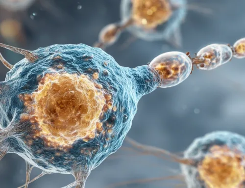
Biomedical Applications
In biomedicine, graphene illustration is used to visualize its interactions with biological systems, including cells, tissues, and molecules. 3D modeling depicts graphene-based drug carriers, biosensors, and scaffolds for tissue engineering, and enables a better understanding of the drug release behavior.
-
Environmental Applications and Catalysts
In these applications, visualization focuses on the surface active sites, adsorption mechanisms, and interactions between graphene, other active materials, and pollutant molecules. These illustrations help scientists understand the mechanism of pollutant removal in a better way.
Cover Arts and Graphene Illustration in 3D
Graphene is an interesting material for energy storage and battery development, due to its high thermochemical stability and electrical conductivity. In the following cover art, graphene was used as a base material in a battery anode. Graphene was exhibited in grey to represent carbon, and silicon nanostructures were demonstrated as blue spheres. Electrons were shown in a vivid, shiny orange to show the electron flow in the graphene structure. Also, Li ions, exhibited as bubble-like structures, are only present in the liquid to exhibit the role of the liquid in charge transfer.
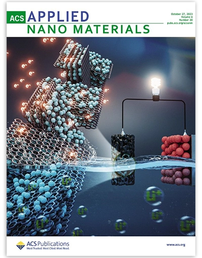
Cover Created by Inmywork Studio (10.1021/acsanm.3c02623)
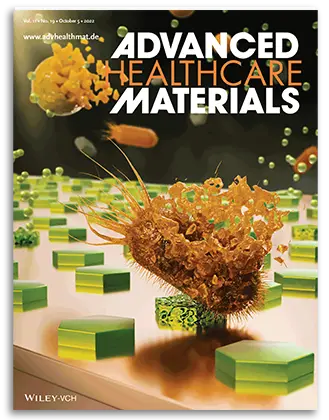
The cover below has a cartoon-like illustrative style, emphasizing the role of functionalized reduced graphene oxide in water treatment. Reduced graphene oxide, exhibited in black, was presented in an eye-catching perspective to show its importance in the work. Functional groups were presented as green spheres and placed on top of the reduced graphene oxide sheet. As it is shown, after sunlight irradiation, the prepared sheets are able to safely degrade Ciprofloxacin, a widely used antibiotic, from water resources.
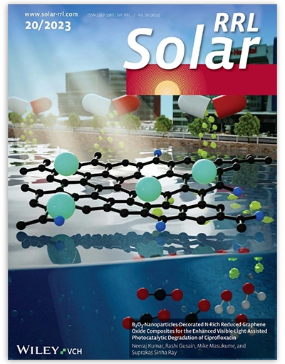
Cover Created by Inmywork Studio (10.1002/solr.202300475)
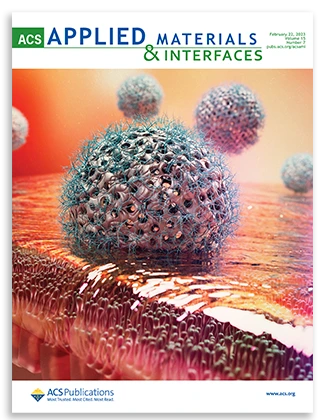
The cover below shows the biocompatibility of dispersible pristine graphene and graphene oxide using a close-to-human animal model. In this cover, the animal model is designed to be held by a scientist, emphasizing the close-to-human concept. Graphene sheets have been displayed under a rainbow light, showing their light-responsivity. As can be seen, the animal model is healthy and being held behind the bottle of graphene, showing the biocompatibility of the graphene.

Cover Created by Inmywork Studio (10.1002/adhm.202570058)
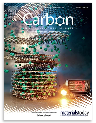
In the field of energy storage, scientists developed a clever network with vertical-porous nanostructures using graphene sheets. The cover below shows graphene layers forming a vertical structure that is both thick and dense and has a high capacity for energy storage. The battery at the back indicates the importance of this structure in the future generation of condensed high-capacity batteries.
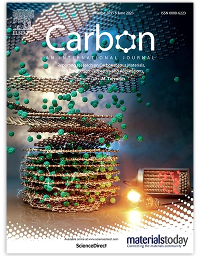
Cover Created by Inmywork Studio (10.1016/j.carbon.2023.02.015)
In the following cover art, a graphene-based carbon-allotropic surface functionalized with EDE-1 in introduced. This graphene-based structure is used for eco-friendly degradation of genetic materials in hospital waste, under UV irradiation and heat exposure. We can see the genetic waste in vivid yellow and red colors, being degraded after exposure to the graphene-based structure and UV light. Since the reaction takes place in water, we can see that the graphene-based structure and genetic waste are shown in an aquatic medium.

Cover Created by Inmywork Studio (10.1039/D3CC03353H)
How can I design a graphene in 3D?
If you need a professional graphene illustration , you can contact Inmywork and use our services. Also, scientists and students can download free 3D scientific bundles like 3D models of Layers or 3D models of Nano Plates and arrange them in Adobe Stager or Blender.
Recent Journal Cover Designs


Leave a Reply