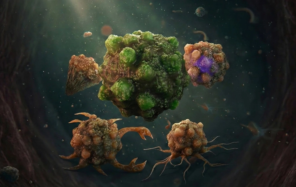
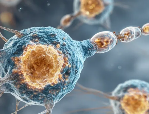
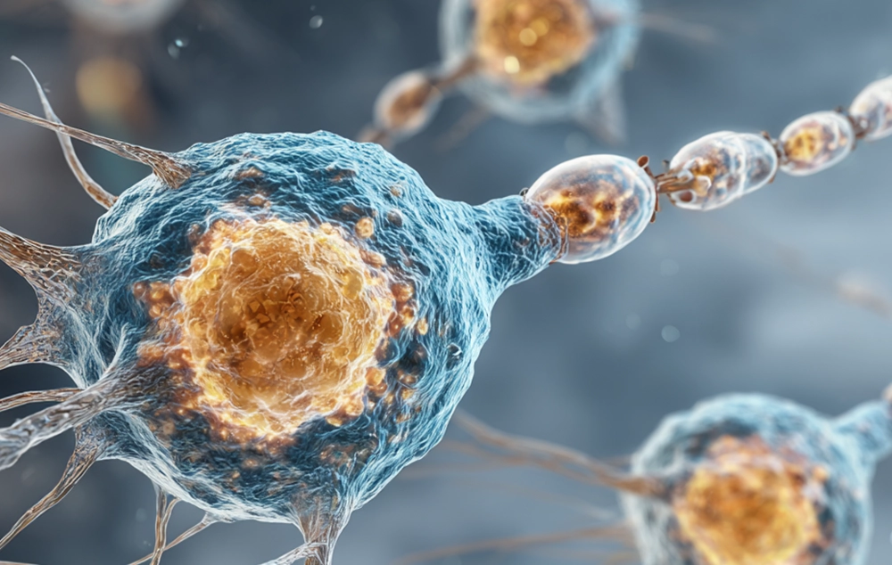
Cancer cells are characterized as cells that lose the ability to regulate their cell cycle and differentiation processes. These cells proliferate uncontrollably, invade surrounding tissues, and may metastasize to other parts of the body through the bloodstream or lymphatic system. They typically display features such as uncontrolled growth, lack of differentiation, and the ability to escape the immune system. Recognizing and visualizing these key features helps communicate their unique behavior effectively.
3D models of cancer cells and their illustrations are valuable in scientific papers and journals for clarifying concepts to audiences, as well as in medical and biological textbooks for visual education. They are also used in scientific presentations and conferences, medical advertisements, health infographics, posters, and websites to enhance understanding and comprehension. Additionally, they are employed in educational and scientific videos to depict the dynamic behavior of these cells.
3D models of Cancer Cell: Visual Features and Symbols
Cancer cell illustrations go beyond mere artistic representation, serving as a bridge between biological reality and communicating complex concepts. Since cancer cells originate from various tissues, they can differ in shape. However, there are common features and widely recognized patterns that serve as known symbols in cancer cell depictions.
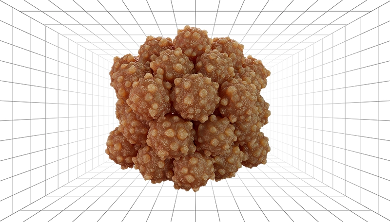
1) Rough and Warty Surface
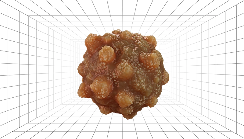
Cells with bulbous protrusions and uneven surfaces are used to illustrate abnormal growth and shape changes. This appearance, inspired by real microscopic images, evokes a sense of danger and threat, making it useful for educating about the morphology of malignant cells.
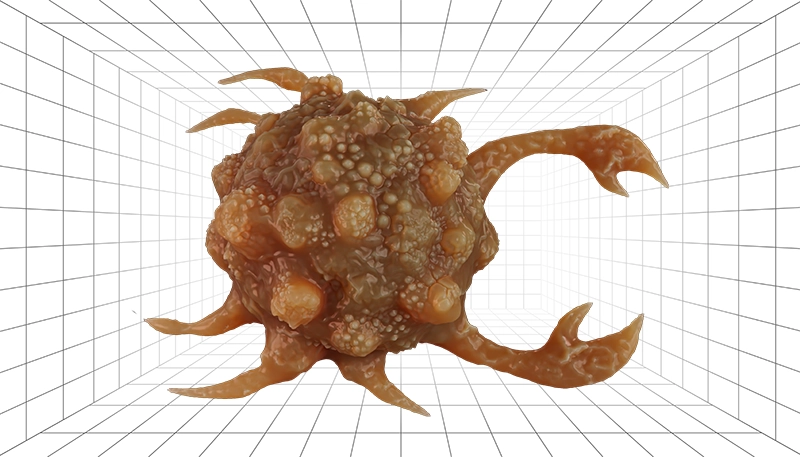
2) Spreading Tendrils
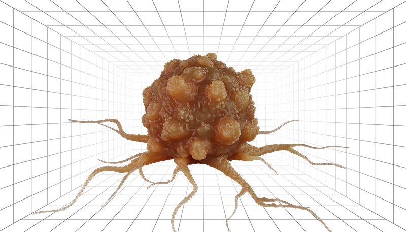
This illustration draws inspiration from the invasive nature of cancer cells and their ability to infiltrate surrounding tissues. Cancer cells are often shown as spherical with outstretched, string-like extensions resembling tree roots or octopus tentacles, conveying a sense of uncontrolled growth. These visuals are effective for explaining invasion and metastasis. For instance, in cancers like sarcoma, invasive cells may be shown with an elongated shape and one or two long extensions. This model is particularly used for cancers affecting connective tissues.
3) Distinctive Nucleus and Shape
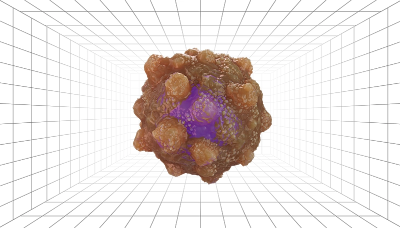
A large nucleus and irregular membrane are hallmark morphological features of cancer cells. The oversized nucleus reflects the cell’s uncontrolled growth and excessive division. Typically, these cells are shown as round with an uneven, bumpy surface and a transparent view of the nucleus. This representation is commonly used to highlight the differences between normal and cancer cells at a microscopic level.
4) Iconic Cancer Symbol
The cancer symbol derives from the word’s origin, displaying a cell shaped like a crab, grasping nearby normal cells with its claws. This imagery is widely used in public visual communication, awareness campaigns, and medical advertisements.
5) Toxic Colors
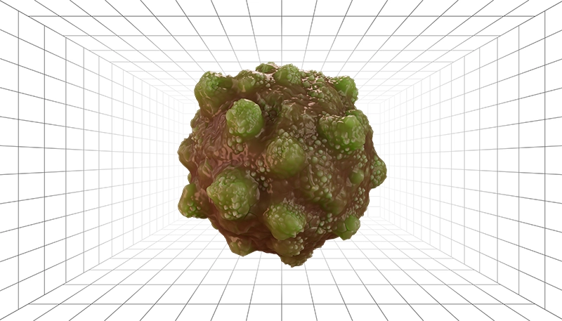
Using colors like black, toxic green, dark red, or purple evokes a sense of toxicity, danger, and disease. These colors are commonly applied in infographics, educational materials, or warning visuals to highlight the threatening nature of cancer cells. In contrast, representations based on diagnostic tests, such as fluorescence or nanomarkers, distinguish normal cells from cancer cells. These cells are often shown with a glowing halo or vibrant colors, used in graphics related to medical imaging or diagnostic techniques.
6) HeLa Cells
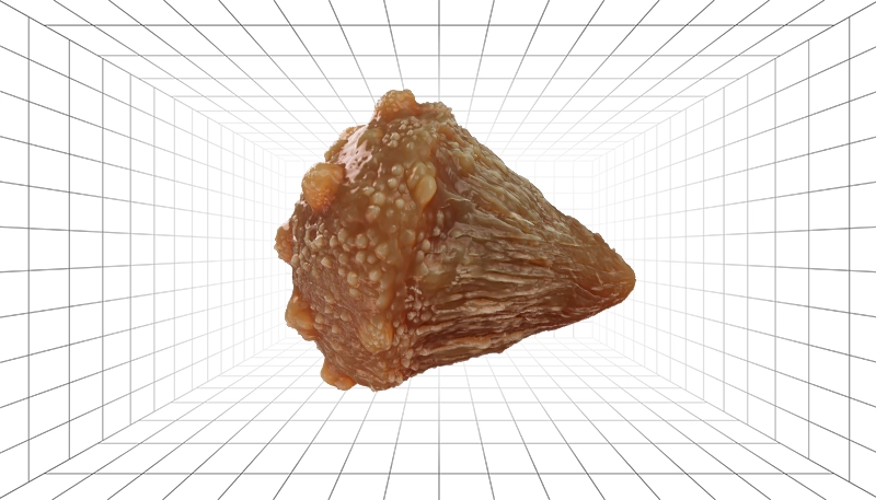
HeLa cells, the most famous cancer cells in research, are known for their stretched, triangular, or conical shapes. As a symbol of cancer research in laboratories, they are frequently modeled in scientific and educational publications to illustrate cell models.
7) Rapid Division
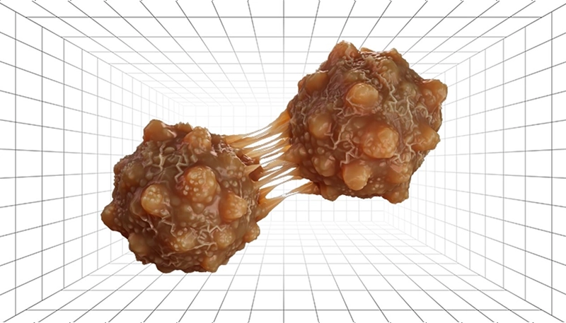
Another model depicts two or more cells undergoing fast, uncontrolled division, often with graphical effects to convey speed, repetition, or rampant mitosis. These visuals are effective in educational presentations to explain cancer cell division and growth.
8) Cell Death: Apoptosis and Necrosis
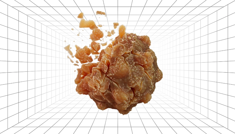
Dying cancer cells are often depicted in two distinct ways. In apoptosis, the cell appears to break apart into small particles, illustrating programmed cell death. In necrosis, the cell is shown breaking apart with larger fragments and debris, representing a more chaotic destruction. These visuals are used to explain concepts like cell death and cancer treatment mechanisms.
9) Tumor Masses
Tumors are clusters of similar cells in a dense formation, varying in size from small (a few cells) to medium or large. These cell masses can be exhibited with cells breaking away and migrating to surrounding areas, effectively illustrating metastasis and cancer spread mechanisms. Additionally, tumors may be shown as a mix of cancer cells with different colors or shapes, useful for studying tumor heterogeneity and drug resistance. For example, brain tumors in neuro-oncology research are often represented with multifaceted, irregular cells, while common cancers like carcinomas are depicted with round cells. Showing tumors near blood vessels or interacting with them is also effective for explaining angiogenesis and nutrient uptake pathways.
10 ) Cancer and the Immune System
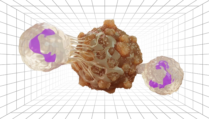
Another common depiction shows immune system components, such as lymphocytes, attacking cancer cells or tumors, or proteins, antibodies, and nanoparticles interacting with them. These visuals are valuable for illustrating immunotherapy, therapeutic biotechnologies, and targeted drug delivery.
Who Benefits from Cancer Cell Illustrations
Cancer cell illustrations are powerful tools that help various fields visualize complex processes, making them easier to understand and study. Below are key areas that benefit from these visuals:
-
Drug Design and Delivery
Illustrations show how nanoparticles or drug molecules interact with cancer cells, highlighting mechanisms of drug action. These visuals help researchers and medical professionals understand how treatments target and destroy cancer cells.
-
Physical Cancer Treatments
Visuals show the effects of treatments like laser or plasma on cancer cell membranes, illustrating how these methods lead to cell death. Such representations are valuable for explaining the impact of physical therapies.
-
Genetic and Stem Cell Research
Illustrations display signaling pathways, genetic mutations, and molecular interactions in cancer cells. These visuals aid in studying the role of genetics and stem cells in cancer development and treatment.
-
Cancer Statistics and Epidemiology
Visualized graphs and diagrams illustrate cancer cell growth within the body or across patient populations. These tools are essential for statistical, clinical, and epidemiological studies, making complex data more accessible.
-
Immunotherapy and Targeted Therapies
Illustrations show how the immune system, antibodies, or targeted treatments attack cancer cells. These visuals are crucial for explaining immunotherapy and precision medicine approaches.
Cancer Cell Journal Covers: Stunning Scientific Visuals
These covers bring to life the beauty and science of cancer cell research through eye-catching designs. Each image highlights key discoveries and concepts, making complex ideas easier to grasp.
Macromolecular Materials and Engineering (Volume 310, Number 2, February 2025):
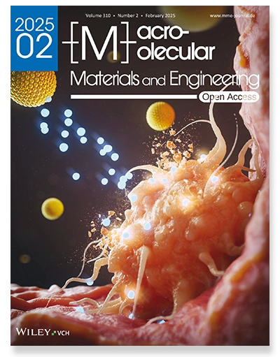
(Cover Created by Inmywork Studio)
The first cover exhibits a dynamic and colorful design that brings cancer cell research to life. The central focus is a glowing, orange tumor cell with tentacle-like extensions, surrounded by bursts of light and cell fragments, showing its active and aggressive nature. Yellow spherical structures, representing drug particles, interact with the cell, releasing energy and breaking it apart, symbolizing a new treatment approach. The artistic use of light and motion highlights how the drug targets and weakens the cancer cell, set against a dark background that emphasizes the vibrant details.
Advanced Healthcare Materials (Volume 14, Number 15, June 10, 2025):

(Cover Created by ABD LAB)
The cover features a striking, vibrant design highlighting cancer treatment in action. A glowing, twisted cancer cell dominates the center, engulfed in a burst of light and particles to reveal its intricate structure. Above, a radiant, disc-shaped symbol and bright yellow spheres depict targeted therapy, blasting the cell with energy and fragments. A nearby DNA strand underscores genetic changes, while the deep red background intensifies the dynamic exchange between the drug and cancer cell.
Advanced Materials (Volume 36, Number 19, May 9, 2024):
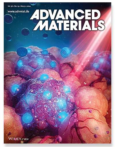
(Cover Created by Inmywork Studio)
The cover presents a striking, high-contrast depiction of cancer cells in motion. The image displays a mass of luminous, irregular, purple cancer cells, encircled by smaller, sharp-edged blue spheres that depict treatment agents. A vivid red laser slices through the scene, representing targeted therapy that shatters the cells, releasing bursts of energy. The vibrant interplay of purple, blue, and red against a deep background dramatizes the confrontation between drug and cancer cells, bringing the science to life.
Cancer Cell (Volume 42, Number 11, November 11, 2024):
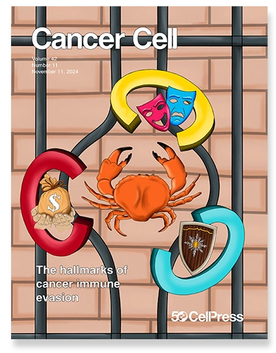
(Cover Created by Galluzzi and colleagues)
This cover features a striking design that reflects a new way to understand how cancer cells escape the immune system. The image shows a crab with claws, symbolizing cancer, surrounded by colorful elements such as masks, a shield, and a money bag, each tied to a unique strategy. The masks represent “camouflage,” where cancer cells hide from the immune system to avoid detection. The shield symbolizes “cytoprotection,” illustrating how these cells protect themselves from harmful treatments. The money bag with the letter “C” hints at “coercion,” where cancer cells weaken the immune response. Together, these visuals illustrate the “three Cs” framework, explaining how cancer resists immunity and treatments in different cases.
How can I design a cancer cell?
3D models of cancer cells and their illustrations play a vital role in conveying scientific, educational, research, and medical marketing concepts. Beyond their visual appeal, these illustrations enhance understanding of cancer characteristics, behaviors, and treatment processes.
On Inmywork Studio, we have a skilled team of creative graphic designers, professional illustrators, and experienced scientific analysts ready to bring your vision to life. Whether you need compelling visualizations of cancer cells or related concepts in 3D, our team can craft accurate and engaging graphics tailored to your needs, perfect for education, research, or awareness campaigns.
Recent Journal Cover Designs


Leave a Reply