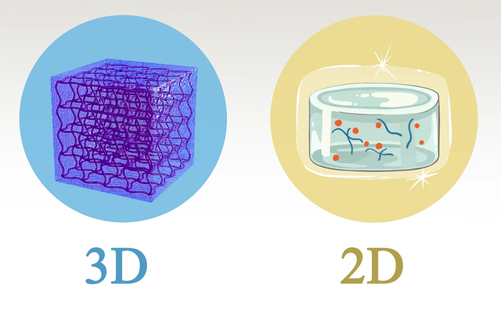
Hydrogels are 3-dimensional polymeric networks with high potential for many different applications, specifically in modern medicine. These structures are able to absorb and contain water up to 95% of their initial weight. Hydrogels can also retain different cargos such as drugs, macromolecules, and nanomaterials. Therefore, they are perfect candidates for drug delivery as well as biosensing, tissue engineering, and many other applications. In this regard, accurate visualization of hydrogel structure is critical for scientists. A good illustration of the hydrogel is crucial for a better understanding of the behavior and the function of the hydrogel.
Hydrogel illustration methods can generally be divided into two categories: experimental illustration (based on microscopy) and digital (based on graphics).
Digital modeling methods for hydrogel illustration
Digital visualization provides flexible and non-invasive ways for hydrogel illustration and simulation. These illustrative methods are widely used in scientific publications, modelling, and presentations. These methods are applicable for 3D illustration and 2D schematics.
Different shapes of hydrogels:
In scientific illustration, hydrogels are mainly represented using simplified symbols, depending on their scale, purpose, and application context. Common representations of hydrogels are as follows:
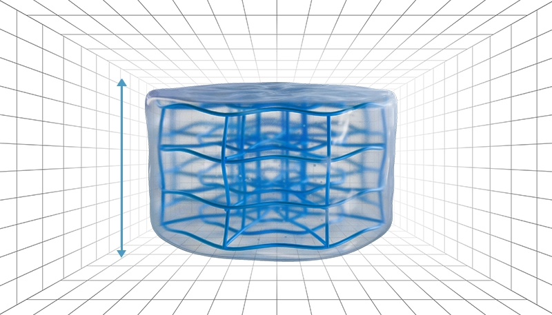
1. Cubes:

These symbols represent solid-like hydrogel structures and are often used in the context of tissue engineering. This type of illustration is usually used when we need to exhibit the polymeric network within the hydrogel structure.
2. Cylinders:
These symbols are typically used to indicate that hydrogels are flexible or can change shape. For instance, showing a hydrogel under pressure or contraction, cylinder shapes can convey this purpose ideally.
3. Spheres or droplets:
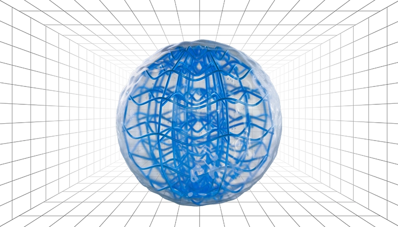
These symbols usually represent microspheres or injectable hydrogels. This type of exhibition is mainly used when we aim to show the hydrogel being injected or dripping from a syringe.
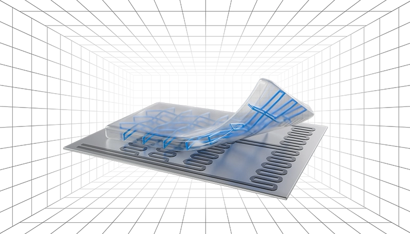
4. Thin layer:
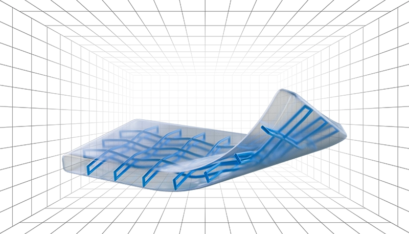
These symbols are commonly used in medical and sensor applications and can indicate bending. When a device component or complex texture involves a hydrogel, layer symbols are the most suitable option.
Different polymer network patterns of hydrogels:
The polymer network pattern within the hydrogel structure also needs simplification in visualization. These chains are mainly shown with lines, meshes, or thread-like textures.
1. Disordered and random form
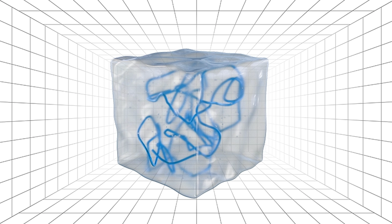
Disordered and random formation of polymers inside the hydrogel represents isotropic hydrogels.
2. Aligned form
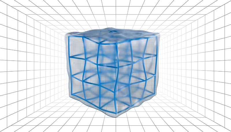
Aligned fibers usually show a cross-linked network, which is often stimuli-responsive.
3. Zoomed-in views
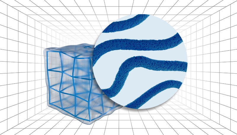
We can also have zoomed-in views to emphasize the porosity and exhibit the texture of the hydrogel or nanostructures.
Key research areas in which hydrogel’s figures are widely applied
- Drug delivery systems
Hydrogels are widely used in targeted and controlled drug release. A good illustration can show how the hydrogel is prepared, loaded with drugs, and releases the drug at specific sites within the body.
- Environmental remediation and pollutant removal
Hydrogels are capable of absorbing pollutants from water and soil. Visual representation highlights the absorption procedure and the interaction of the hydrogel network with the pollutant.
- Smart bandages, wound healing, and medical care
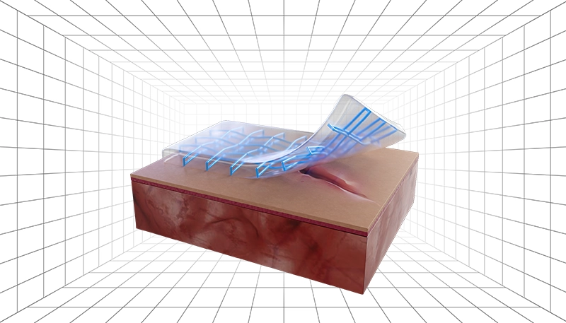
Advanced wound dressing is one of the most important applications of hydrogels, since they maintain moisture and can be tailored to exhibit antibacterial activity. Illustrative methods can help show how the hydrogel helps with wound closure and resists bacteria.
- Biosensing and responsive hydrogel systems
Stimuli responsive hydrogels react to pH, temperature, or specific enzymes for biosensing applications. Diagrams and figures can help explain the sensing mechanism and signal appearance process.
- Tissue engineering and regenerative medicine
Hydrogels serve as scaffolds in tissue engineering and can support cell growth. Illustrative methods can help show how cells interact with the hydrogel matrix and how the structure supports tissue formation.
Samples of hydrogels’ illustration on cover pages
Hydrogels are highly studied for 3D printing of scaffolds, which will be used in a variety of applications, especially tissue engineering. Scaffolds in tissue engineering are designed and prepared with similarity to the target tissue, so they can effectively mimic it in a biological environment.
Biomaterials Science (21 July 2025, Issue 14)

(Cover Created by Inmywork Studio)
The sample cover shows 3D printing of a hydrogel for dental tissue engineering. The hydrogel is displayed as a yellow gel structure, being printed into the tooth and forming a scaffold. Bacteria near the filled tooth are being exploded due to the antibacterial activity of the hydrogel.
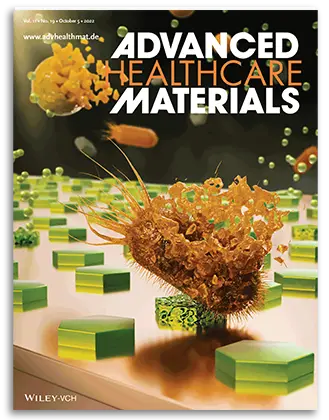
Advanced Healthcare Materials (Volume 12, Issue 18, July 17, 2023)
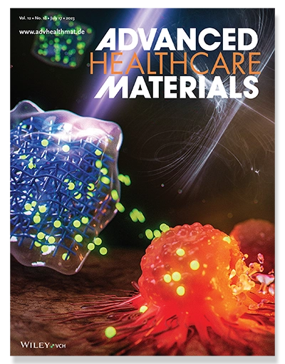
(Cover Created by Inmywork Studio)
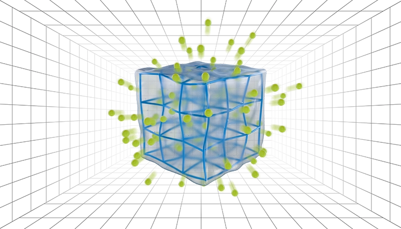
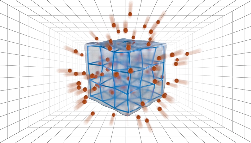
Hydrogels are great candidates for cancer therapy applications due to their stimuli-responsiveness and on-demand drug release ability. As shown in the cover image below, hydrogels can absorb the desired drug molecule and release it under specific stimuli in the tumor location. In this cover, hydrogel is illustrated as a cube with a clear color that we can see the polymeric network inside. This composite contains drug molecules, which are displayed with a phosphorous bright green color. The drug molecules are released under laser radiation to destroy cancerous cells at the tumor site. We can clearly see the destruction of the cancer cell, illustrated in a bright orange color.
ACS Applied Materials & Interfaces (2024. Volume 16, Issue 18)

(Cover Created by Inmywork Studio)
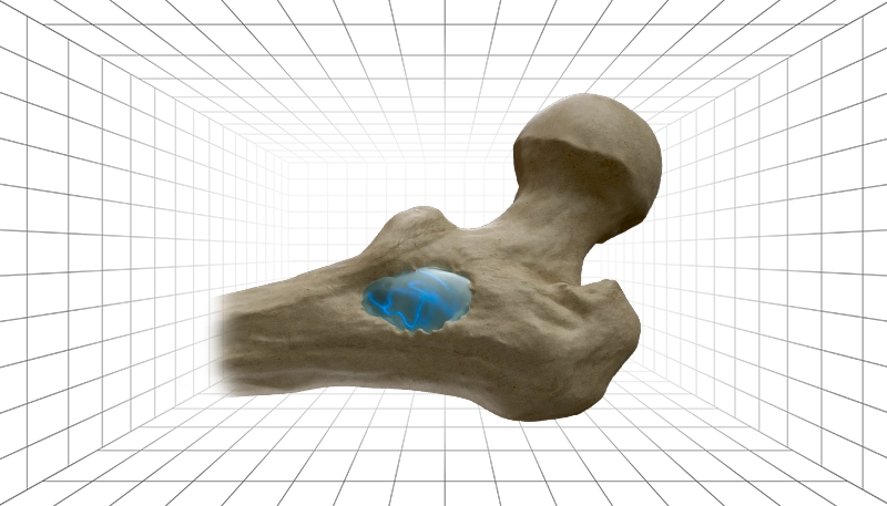
The sample cover below shows how biocomposites based on hydrogels can be used for bone tissue engineering. The biocomposite is exhibited as a porous dark filler in the defected bone, exhibiting anticancer and antibacterial activity under laser irradiation. As we can see, the cancerous cells and bacteria near the laser are being destroyed.
Advanced Materials Interfaces (Volume 12, Issue 15, August 7, 2025)
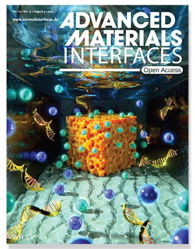
(Cover Created by Inmywork Studio)
The following cover image aims to show the ion concentration polarization near a hydrogel within a nanofluidic system. The hydrogel is illustrated as a 3D porous cube with a vivid yellow color at the far side of the tank. Ions, displayed as bright blue and purple spheres, and RNA are floating around the nanofluidic system. The design shows that the concentration of a certain type of ions is higher around the hydrogel, emphasizing the hydrogel’s selectivity and potential in biosensing.
Small (Volume 20, Issue 8, February 22, 2024)
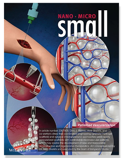
(Cover Created by Inmywork Studio)
The presented cover image, shows how bulk hydrogel spheres can be used for revascularization and soft tissue repair. A combination of micropuncture and granular hydrogel scaffold was used to create vascularized scaffolds. Hand represents the chosen soft tissue, and hydrogel, exhibited as white spheres dripping from the needle, fills up the spaces between vessels to promote tissue repair.
How can I design a hydrogel?
Hydrogels are outstanding polymeric materials and have attracted attention due to their tunable characteristics. The use of hydrogels is growing in a variety of scientific fields, such as cancer therapy, tissue engineering, regenerative medicine, environmental remediation, and biosensing. If you need a professional hydrogel illustration , you can contact Inmywork.
Recent Journal Cover Designs


Leave a Reply